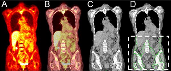
Figure 1.

(A) PET, (B) fused PET/CT, and (C) CT coronal section of a representative patient. The segmented skeletal muscle is outlined by green lines and overlaid onto the corresponding CT coronal section (D). The dotted white lines in (D) show the standardized body region used for segmentation in our study.
