. 2018 Apr 24;9(18):4299–4307. doi: 10.1039/c8sc00428e

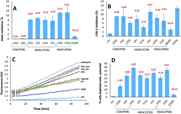
Fig. 5. (A) HDAC inhibition. PSN-1 cells were incubated for 24 h with 1 μM of tested complexes. The HDAC activity was determined in cells by FLUOR DE LYS® HDAC fluorometric activity assay kit (Enzo Life Sciences) following the manufacturers' instructions. In all cases, data are the means of at least three independent experiments. (B) COX-2 expression. PSN-1 cells were incubated for 24 h with 1 μM of tested complexes. The inhibition of COX-2 was measured by using COX activity assay kit (Cayman Chemical, Ann Arbor, USA) following the manufacturers' instructions. In all cases, data are the means of at least three independent experiments. Error bars indicate SD. (C) ROS production. PSN-1 cells were preincubated in PBS/10 mM glucose medium for 20 min at 37 °C in the presence of 10 μM CM–DCFDA and then treated with equimolar doses (10 μM) of Pt(iv) derivatives. The fluorescence of DCF was measured. In all cases, data are the means of three independent experiments. (D) Effects on cellular mitochondrial membrane potential. PSN-1 cells were treated for 24 h with 1 μM of tested compounds. The percentage of cells with hypopolarized mitochondrial membrane potential was determined by Mito-ID® Membrane Potential Kit. The IC50 values of the compounds against the PSN-1 cells are depicted in red.

Fig. 5