. 2018 Apr 16;10(16):7769–7779. doi: 10.1039/c8nr00196k

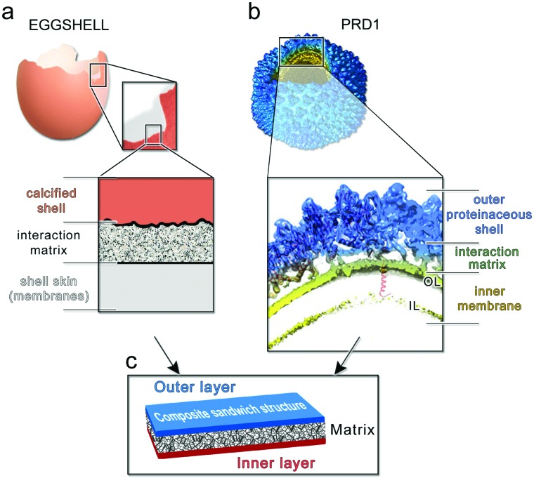
Fig. 6. (a) Multilayered structure of an avian eggshell; (b) cryo-electron density of wt PRD1 with an octant removed displayed in Chimera45 to show (inset) the layered/composite structure of the virion with the proteinaceous shell in blue, the membrane-vesicle in yellow-lime (OL: outer leaflet; IL: inner leaflet) and with the horizontal black lines marking the distinct layers and the interacting matrix region between the capsid and the membrane. As cartoon representation in red fitted in density the P3 MCPs with N-terminal α-helices interacting with the OL and the P16 transmembrane protein crossing the membrane vesicle. The dsDNA has been removed from within the membrane vesicle for clarity; (c) schematic of a composite sandwich material to which (a) and (b) recapitulate.

Fig. 6