Figure 1.

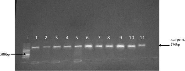
Gel electrophoresis of PCR amplification of nuc gene. L = 100 bp ladder 1, 2, 3, 4, 5, 7, 8, 9, 10, 11 = nuc positive isolates; 6 = nuc positive control; not labeled = nuc negative control (PCR water)

Gel electrophoresis of PCR amplification of nuc gene. L = 100 bp ladder 1, 2, 3, 4, 5, 7, 8, 9, 10, 11 = nuc positive isolates; 6 = nuc positive control; not labeled = nuc negative control (PCR water)