. 2018 May 1;10(18):8413–8421. doi: 10.1039/c8nr00603b

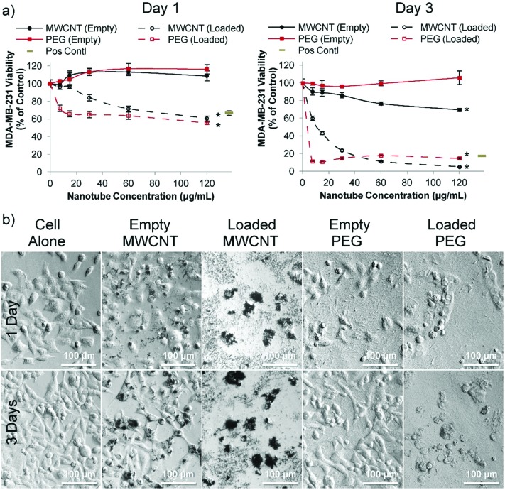
Fig. 3. Cytotoxicity of doxorubicin loaded nanotubes with MDA-MB-231 breast cancer cells in vitro. (a) After one day (left hand graph), MDA-MB-231 cells incubated with either doxorubicin loaded MWCNTs or doxorubicin loaded PEG nanotubes show a significant decrease in viability which is further reduced by day three (as measured via the PrestoBlue assay). All nanotubes were subject to the same loading solutions (80 μg of doxorubicin per 1 mg of nanotubes (0.08 : 1 ratio)). Positive control is doxorubicin in solution at a concentration of 4 μg mL–1 (b) corresponding light microscope images of the 60 μg mL–1 nanotube concentration show the effect of the loaded nanotubes on cell morphology (n = 4), error bars represent ± standard deviation, * represents statistical significant difference to empty PEG nanotubes, (two way ANOVA with Tukey's multiple comparison test (P ≤ 0.05)).

Fig. 3