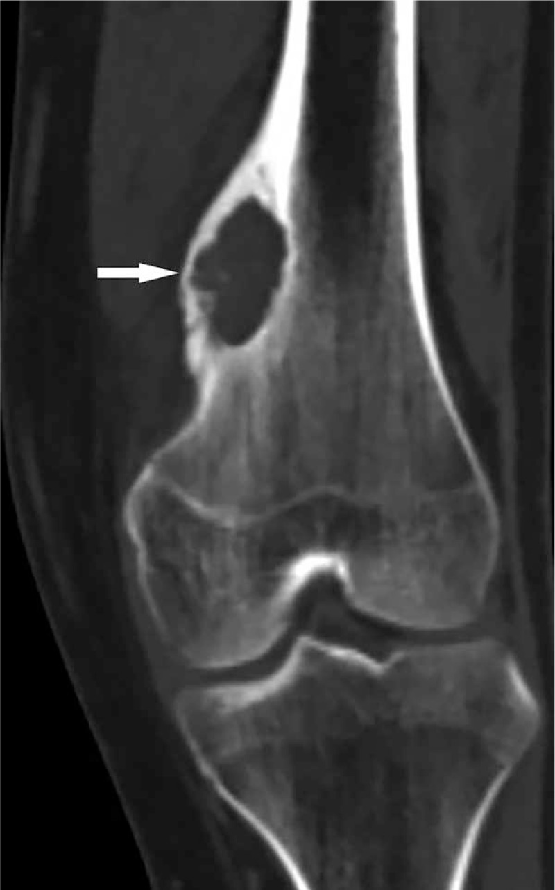
Figure 3.

Coronary reconstruction CT images show the local cortical thickening, and lytic bone destruction of medial-distal left femoral metaphysis (white arrow) with internally, stippled calcification, and marginal sclerosis. CT = computed tomography.
