
Figure 1.
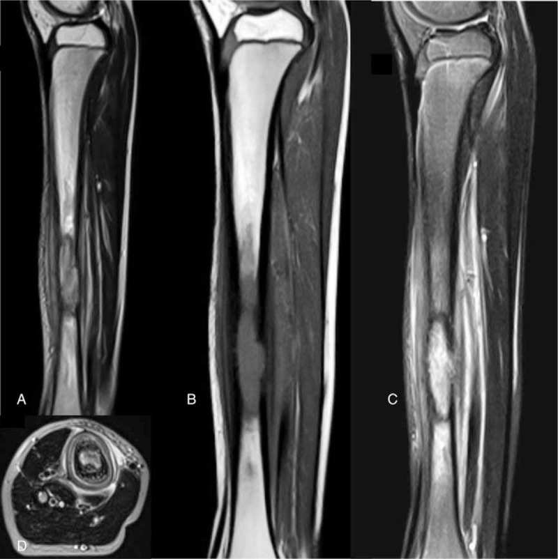
T2WI, T1WI, and STIR MR sequences of the right tibia of an 8-year-old boy with Langerhans cell histiocytosis. The ovoid lesion with isointense and hypointense signals on T1 sequence (A) and hyperintense signals on T2 sequence (B) were detected in the lower 1/3 of the medullary cavity of the right tibia. The lesion had a sclerotic margin, which showed hyperintense signals on STIR sequence (C). There was also a layered periosteal reaction (D). MR = magnetic resonance, STIR = short time inversion recovery sequences.