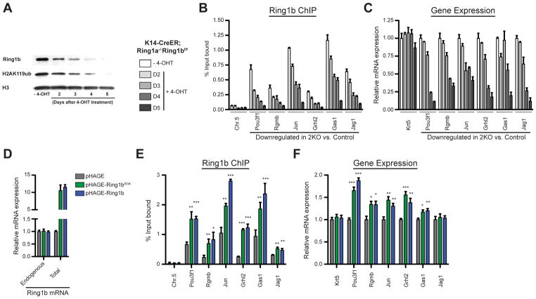
Figure 5. Ring1b binding positively regulates gene expression.
(A–C) PRC1 loss-of-function analyses in K14-CreER; Ring1a−/− Ring1bflox/flox epidermal progenitor culture upon 4-OHT treatment. (A) Western blot showing changes in Ring1b and H2AK119ub protein levels over time. (B) ChIP-qPCR showing changes in Ring1b binding. Data are mean ±SEM, n=3. (C) RT-qPCR analysis of changes in the expression of Ring1b target genes. Data are mean ±SEM, n=3. (D–F) Ring1b overexpression analyses in wild type epidermal progenitor culture. (D) RT-qPCR analysis of endogenous and total Ring1b mRNA. (E) ChIP-qPCR showing changes in Ring1b binding. n=3. (F) RT-qPCR analysis of changes in expression of Ring1b target genes. n=5. Data in graphs (E and F) are mean ±SEM. *p<0.05; **p<0.01; ***p<0.001 (two-sided t test). See also Figure S6.

