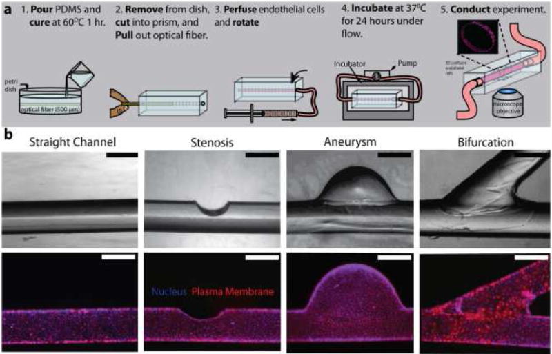
Fig. 3. Endothelialized microfluidics can be developed using off-the-shelf laboratory materials.

a) Fabrication process flow of this “do-it-yourself” endothelialized microfluidic device. b) Different microvascular geometries created via slight alterations in the fabrication protocol. Scale bars represent 500 μm. Image reproduced, with permission, from reference 28.
