
Fig.4.
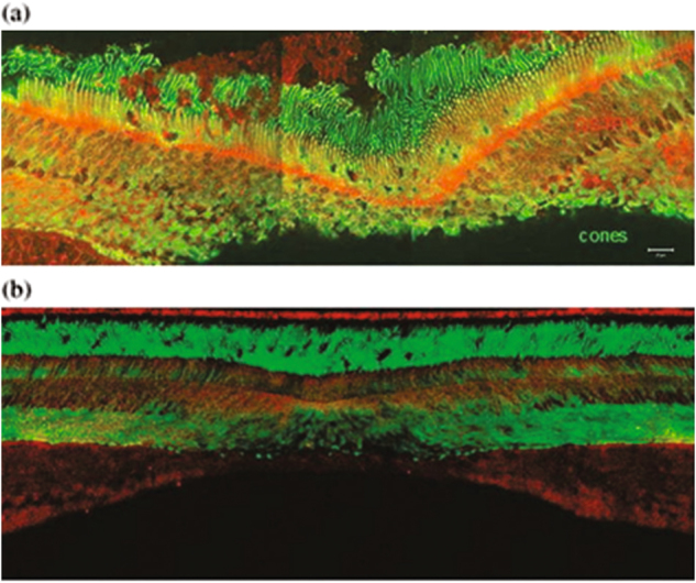
The retinal distribution of macular pigment binding proteins. a GSTP1 labeling of foveal cones in the macula of a 3-year-old monkey. This montage shows strongest labeling by antibody against GSTP1 (red) over the myoid and ellipsoid regions of cones identified by monoclonal antibody (7G6, green). b A low-magnification view of a near-foveal retina section in which N-62 StAR (red) identifies StARD3, an anti-cone arrestin monoclonal antibody (7G6, green) identifies monkey cones. Images courtesy of Dr. Jeanne M. Frederick