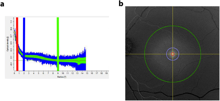
Fig. 7.
Macular pigment readout from an unsupplemented normal control obtained by dual wavelength autofluorescence imaging on a Heidelberg Spectralis. a Macular pigment tracing at 0.5° (red line), 2° (blue line), and 9° (green line). b Autofluorescent image showing the fovea and the degrees (0.5°, red; 2°, blue; 9°, green) from the center of the macula lutea

