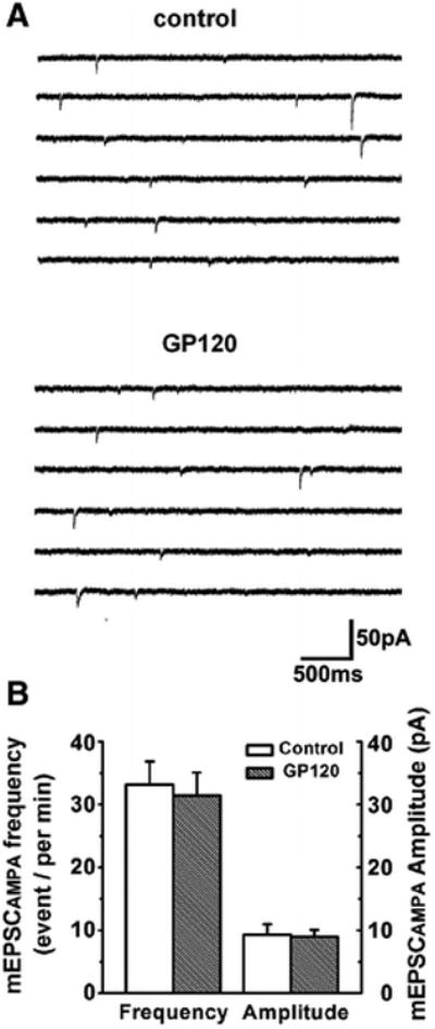
Figure 4. Gp120 failure on alteration of spontaneous mini EPSCAMPAR (mEPSCAMPAR).

A. Representative traces of mEPSCAMPAR recorded from a CA1 neuron in a rat hippocampal slice in the presence of NMDA receptor antagonist AP-V (50μM) in the perfusate. Addition of gp120 to the perfusate failed to alter mEPSCAMPAR. B. Bar graph showing average event frequency and amplitude of mEPSCAMPAR were not significantly changed after addition og gp120 (200pM) to the perfusate (n=9, p> 0.05 vs control.
