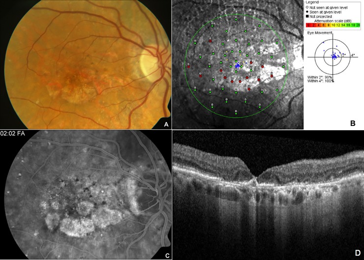
Fig 1.
Representative color fundus photograph (A), SLO macular microperimetry (B), fluorescein angiogram (C) and optical coherence tomography (D) data of the right eye of a patient with bilateral advanced age-related macular degeneration. Color fundus photograph (A) showed patchy area of geographic atrophy with scattered drusen in the macula. SLO macular microperimetry (B) showed good fixation (blue dots) with 2–3 disc diameters sized inferior paracentral scotoma secondary to geographic atrophy sparing the central fovea as indicated by red (non-responding)/green (responding) dots leading to paracentral moderate scotoma (Score 4). Late phase angiogram (C) showed staining patterns along the atrophy area with diffuse area of outer retinal layer irregularity on OCT with geographic atrophy (D).

