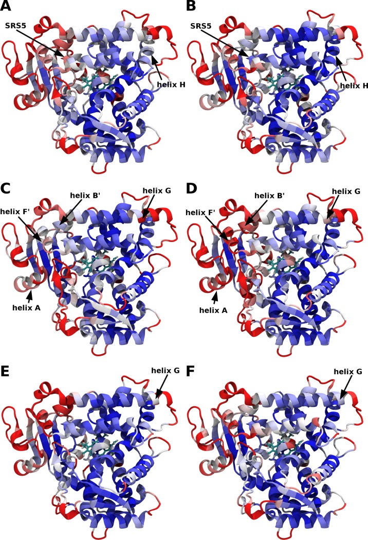
Fig 2. Root Mean Square Fluctuations (RMSF) of the Cα atoms during the MD simulations of CYP2C9.
The RMSF computed for five merged MD trajectories of (A) WT apo, (B) A477T apo, (C) WT diclofenac-bound, (D) A477T diclofenac-bound, (E) WT losartan-bound and (F) A477T losartan-bound are mapped on the CYP2C9 crystallographic structure (PDB ID: 1OG5) [25]. The color code of RMSF is ranging from 0.7 Å (blue) to 1.6 Å (red). The black arrows show noticeable differences between the WT and the mutant RMSF.

