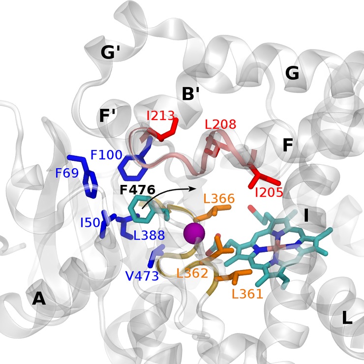
Fig 4. Hydrophobic contacts of F476 and other residues of the CYP2C9 binding pocket monitored over the MD simulations.
Residues found in contact with F476 are depicted in sticks. Three distinct hydrophobic clusters are found and colored in blue, red and orange. The purple sphere represents the location of the mutation.

