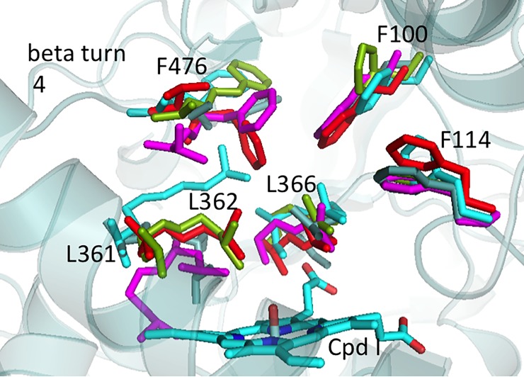
Fig 8. Key residues of the active site of the best identified CYP2C9 structures for substrate docking generated from MD simulations of diclofenac-bound CYP2C9.
The side chains of the diclofenac-bound WT structures are colored in grey (centroid 0), cyan (centroid 1), light green (centroid 2). The side chains of the diclofenac-bound A477T structures are colored in red (centroid 0) and violet (centroid 1).

