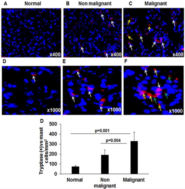
Figure 3. Mast cells analysis in normal, malignant and non-malignant pancreatic patients.

Anti-tryptase immunostaining was performed to detect mast cells in tissue sections and anti-tryptase positive mast cells were shown in a representative photomicrograph of normal (A, D), non-malignant (B, E), and malignant pancreatic tissue sections (C, F). White arrows indicate tryptase positive intact mast cells (A–D); degranulated mast cells (marked by yellow arrows) and extracellular tryptase granules (marked by green arrows) are detected only in malignant pancreatic tissue sections (C, F). The quantitation of mast cell numbers in normal, non-malignant and malignant pancreatic tissue sections were detected and found significantly induced compared to the normal pancreas tissue sections (G). Representative photomicrographs presented as ×400 and ×1000 of original magnification. The quantitative data are expressed as mean ± S.D, n=3 for normal and n = 4 for malignant and n=3 non-malignant patient.
