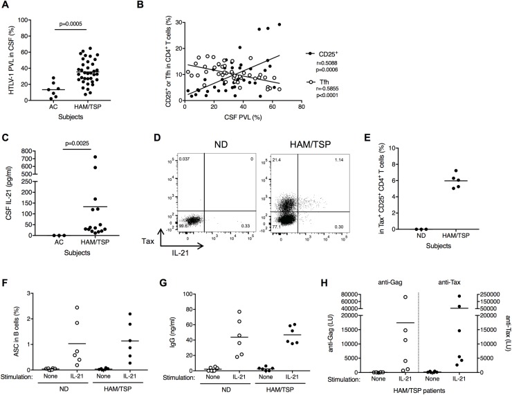
Fig 6. Involvement of CD4+CD25+ T cells with B cell help in CSF of HAM/TSP patients.
(A) Comparison of HTLV-1 PVL in CSF of ACs (n = 7) and HAM/TSP patients (n = 36) using Mann-Whitney Test. (B) Correlation of HTLV-1 PVL with CD4+CD25+ T cells and memory Tfh cells in HTLV-1-infected subjects using Spearman’s rank correlation test. (C) Comparison of IL-21 in CSF of HAM/TSP patients and ACs using unpaired t test. (D) Representative dot plots of IL-21 and Tax staining in CD4+CD25+ T cells of a ND and a HAM/TSP patient after culture for 24 hours without any exogenous stimulation. (E) Detection of IL-21 in Tax-expressing CD4+CD25+ T cells of HAM/TSP patients after culture for 24 hours without any exogenous stimulation. (F) Generation of ASCs subsets in B cells cultured with and without rhIL-21. The data were obtained from cultured B cells of NDs and HAM/TSP patients (n = 6). The horizontal line represents the mean. (G) Detection of human IgG in the B cell culture supernatants of NDs and HAM/TSP patients. (H) Detection of antibodies for HTLV-1 Gag and Tax in the B cell culture supernatants of HAM/TSP patients.

