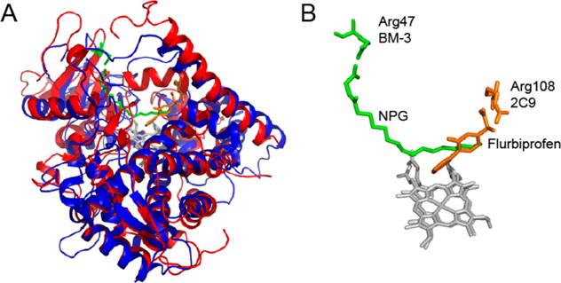
Figure 7.

Comparison of the position of arginine in NPG bound to BMP in 4KPA (4KPA, red and green) and flurbiprofen bound to CYP2C9 (1R90, blue and orange). Both proteins have the same overall fold (A); however, the arginine residues in the binding pocket are in different locations to accommodate the difference in the size of the substrates (B). The structures were prepared in PyMOL.41
