
Figure 2.
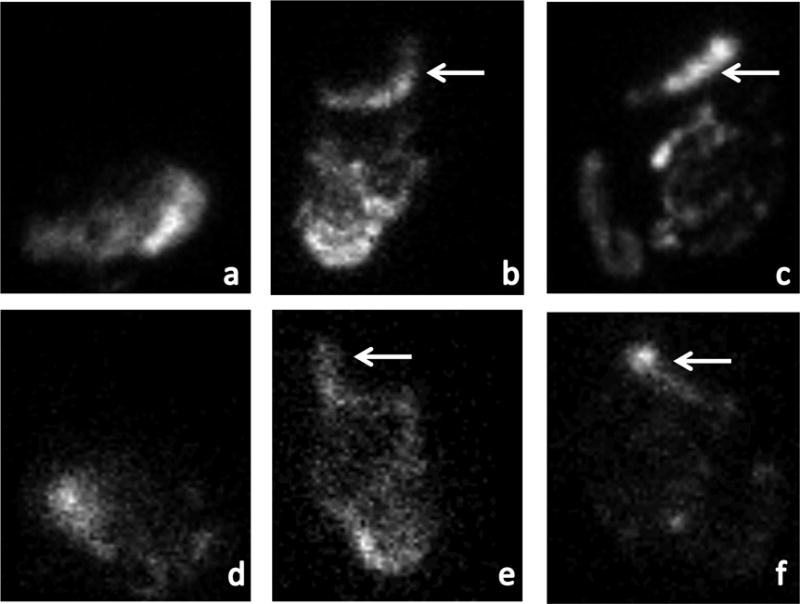
Gastric emptying abnormalities in participants with vagal dysfunction
shows gastric emptying scintigraphy images at 4 hours after ingestion of a radiolabeled meal. (a) is a normal anterior image in which all radiolabeled material has exited the stomach and moved distally. (b) and (c) are anterior images from the participants with vagal dysfunction and gastroparesis demonstrating retained radiolabeled material in the stomach (arrows). (d)-(f) are the analogous posterior images.