
Figure 6.
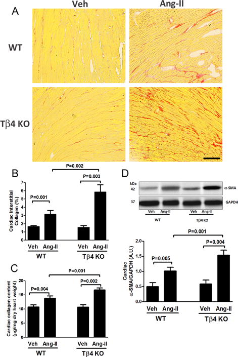
Effect of Ang-II on cardiac fibrosis and α-smooth muscle actin (α-SMA) protein expression in the heart of Tβ4 KO mice. (A) Representative images of cardiac interstitial fibrosis. Red color indicates total collagen content by picrosirius red staining. Images were captured using a 20× microscope objective. Scale bar = 100 μM. In Ang-II infusion, total cardiac collagen content increased significantly in Tβ4 KO mice, compared with that in the WT. (B) Quantitative data for Figure 6A. (C) Total cardiac collagen content was further quantified by a biochemical hydroxyproline assay. (D) Top figure: At baseline, no significant difference in cardiac α-SMA expression was observed; Ang-II infusion significantly increased α-SMA expression in the heart of Tβ4 KO mice, compared with that in WT mice. Bottom figure shows the quantitative data. All data are expressed as the mean ± SEM, n = 6-10 in each group.