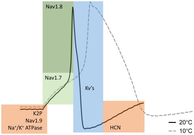
Figure 5. Main molecular components of the action potential.
Shown is the typical shape of an action potential in a mouse DRG neuron at room temperature (black trace) along with major electrical conduits. Colored regions denote the areas of contribution of the indicated channels’ activity to the different phases of the action potential. Nav1.7 contributes to the early depolarization and upstroke (light green). Nav1.8 contributes to the rapid upstroke (dark green). Kv’s repolarize the cell in the falling phase (blue). K2P’s, Nav1.9, and Na+/K+-ATPase regulate the resting potential of the cell, and HCN channels drive the return of the resting potential from the after-hyperpolarization (orange). At cold temperature, the action potential becomes much slower and broader due to temperature dependent changes of the underlying channel activity (dashed gray trace).

