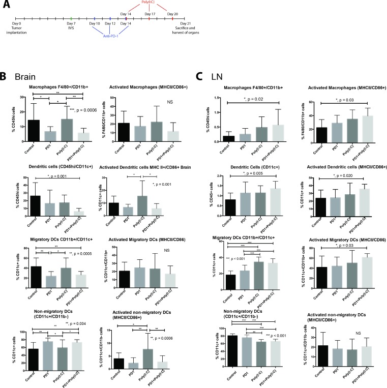
Figure 1. TLR3 agonist enhances activation of dendritic cells (DCs).
(A) Treatment schedule. Mice that received an intracranial injection with GL261-luciferase cells were imaged with bioluminescence (IVIS) on day 7 to ensure similar tumor burden in all mice, after IVIS imaging, mice were randomized to each treatment group. Anti-PD-1 treatment was given on days 10, 12, and 14 via intraperitoneal (i.p.) injection. Poly(I:C) was given on days 14, 17, and 20 via i.p. injection. (B–C) Evaluation of macrophage (F4/80+/CD11b+), Dendritic cells (DC, CD11c+), migratory DCs (CD11c+/CD11b+), and non-migratory DCs (CD11c+/CD11b−) and their activation status in the brain (B) and draining lymph node (C). Bar charts show percentage of cells gating on CD45+ and gating on CD45hi in the brain. We observed an increase in the percentage and activation of macrophages and DCs in deep cervical lymph nodes, particularly in the case of migratory DCs. Interestingly, the percentage of macrophages and DCs infiltrating the brain tumor decreased in the anti-PD-1+poly(I:C) group as compared to control mice. Data are represented as mean ± SEM. All experiments repeated in triplicate with ≥5 mice per arm. P-values were determined by ANOVA, and, *denotes statistical significance (p < 0.05).

