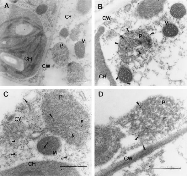
Figure 6.
EM immunocytochemical localization of NADP-ICDH in pea leaves. The electron micrographs are representative of thin sections of pea leaves. Cell sections were probed with preimmune serum (dilution 1:500) (A). Immunogold labeling with anti-pea NADP-ICDH (dilution 1:500) was carried out in young (B) and senescent (C and D) pea leaves. Arrows indicate 15-nm gold particles. CH, Chloroplast; CW, cell wall; M, mitochondrion; P, peroxisome; CY, cytosol. Bars = 0.5 μm.

