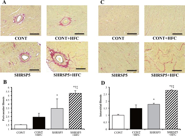
Fig. 4.

Evaluation of cardiac fibrosis in the 4 groups at 18 weeks of age
AD: Collagen deposition as revealed by picro-sirius red (PSR) staining in perivascular (A) and interstitial (C) regions of the LV myocardium. The perivascular fibrosis area was corrected by the area of the perimeter (B), and interstitial fibrosis was expressed relative to the CONT group (D). Scale bars = 100 µm. All data are shown as means ± SE; n = 5 in each group. *P < 0.05 vs. the CONT group, †P < 0.05 vs. the CONT + HFC group, ‡P < 0.05 vs. the SHRSP5 group.
