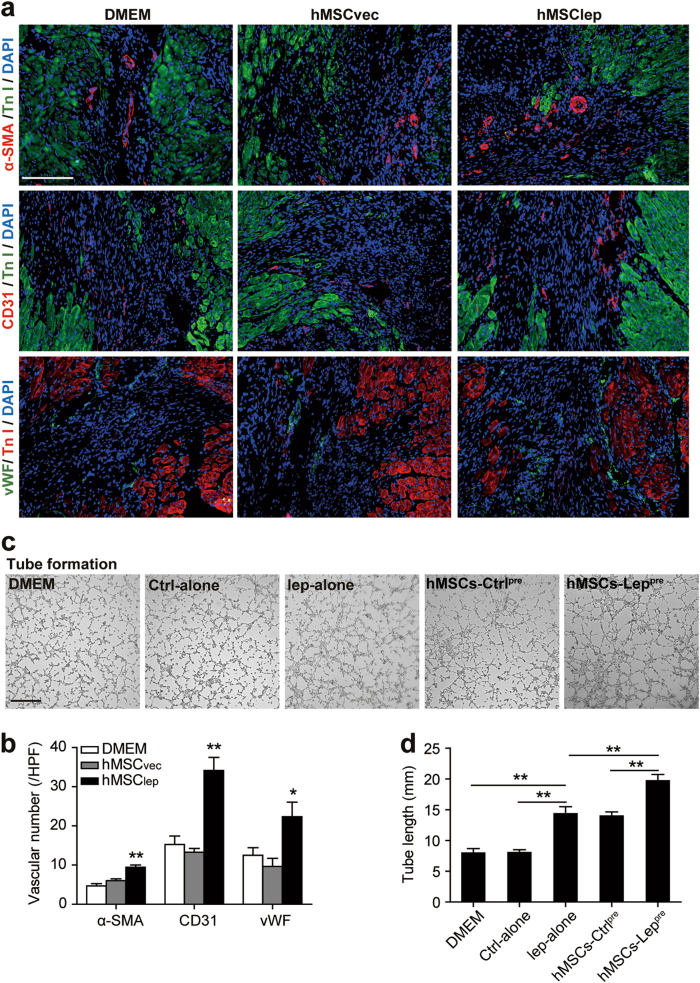
Fig. 3. Paracrine efficacy of leptin-enhanced hMSCs improved angiogenesis.
a, b Representative immunoflourescence of α-SMA, CD31, and vWF in the peri-infarct zone of ischemic hearts using heart tissue slides obtained from DMEM, hMSCvec, and hMSClep group mice (n = 7 for hMSClep group, n = 5 for DMEM, and hMSCvec group each) at day 28 post MI and angiogenesis process was quantified by 6–8 high-power field (HPF) per section in the bar graphs. Scale bar, 100 μm. c, d Tube formation assay was conducted using HUVECs cultured with conditioned medium obtained from DMEM alone, control-alone, leptin alone, hMSCs-Ctrlpre, or hMSCs-Leppre. The conditioned medium had been normalized by an equivalent number of hMSCs (1 × 10⁶ cells). The quantification of tube formation was shown in bar graphs. Scale bar, 50 μm. Independent in vitro experiment was repeated three times. Data were shown as mean ± SEM. *denotes P < 0.05, **P < 0.01

