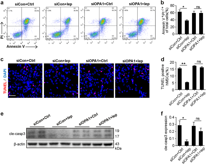
Fig. 5. OPA1 was required for the protective effects of leptin in response to GSDH stress.
a, b Annexin V/PI staining was performed to measure cellular apoptosis and necrosis, and early (Q3) and late (Q2) apoptosis events were quantified simultaneously. c, d Representative images of TUNEL staining and DAPI staining. Scale bar, 50 μm. Quantification of apoptotic cells by TUNEL-positive nuclei. e, f Cleaved caspase 3 (cle-caspase 3) protein expression of WCL was detected by western blot; β-actin served as a loading control. Independent experiments were performed three times. Data were shown as mean ± SEM. *denotes P < 0.05. **P < 0.01

