Figure 3.
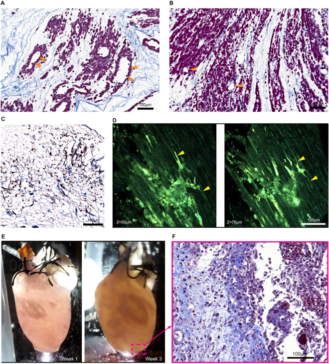
Perfusion recellularization of HEK293 cells, fibroblasts, and h-iPS-CPCs into 4-Flow cannulated hearts. (A) HEK293 cells delivered through only the SV of a 4-Flow heart perfuse through the cardiac veins and throughout the walls of the heart and distribute within the matrix proximal to the vasculature maintaining a hollow lumen (n = 3). (B) HEK293 cells delivered through both SV to the cardiac vein and AA to the coronary arteries are more thoroughly dispersed into the tissue walls leaving the vasculature hollow (orange triangles) (n = 3). (C) Perfusion culture of a primary human cardiac fibroblasts repopulated 4-Flow heart demonstrates cell survival, attachment, and invasion into the tissue (n = 3). Multiphoton imaging captures collagen fiber structures through second harmonic generation seen in cyan while autofluorescence of elastin and cells, and Hoechst nuclear staining is shown in green-yellow. (D) Multiphoton imaging of the 2-week 4-Flow heart reveals the lamellipodia extensions of the fibroblasts (yellow triangles) interacting with the decellularized matrix in three-dimension (n = 3). (E) Macroscopic observation of the recellularized heart with h-iPS-CPCs during the 3 weeks of culture indicate change in opacity (n = 3). (F) Corresponding representative image of Masson’s Trichrome staining shows cells are embedded within the collagen matrix. Histology sections were stained with Masson’s trichrome in which cell nuclei are purple and collagen fibers are blue.

