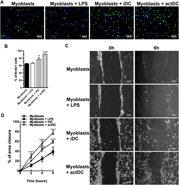
Fig. 4. DCs incubation increase the proliferation and migration of myoblasts.
a Myoblasts were co-cultured with iDC or actDCs for 48 h and the proliferation was evaluated by BrdU incorporation. b Quantitative analysis of the proliferation assay. The data are representative of three experiments. c Images representative of scratched areas from myoblasts alone or after incubation with LPS, or co-cultured with iDC or actDCs for 48 h. d The data are expressed as the percentage of the closed area. Data shown as means ± SE of duplicate wells and are representative of three different experiments. Bars represent 100 μm. *p < 0.05; ***p < 0.001, and ****p < 0.0001 compared to myoblasts

