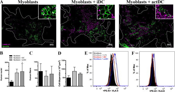
Fig. 6. Co-injection of human myoblasts and DCs into the Tibialis anterior (TA) muscle pre-injured by cryolesion in Rag-/-IL2R-/- mice.
a Immunostaining for human specific lamin A/C (magenta) and spectrin (green) on muscle sections obtained 30 days after cryolesion of the tibialis anterior of Rag−/−IL2R−/− mice. The injections of myoblasts, myoblasts plus iDC or myoblasts plus actDCs were made in 3–4 TA muscles per group. Bars represent 100 μm. The graphs show the number of human nuclei (b), human spectrin postive muscle fibers (c), and cell dispersion (d). The data represent one experiment. Myoblasts were seeded in co-culture with iDC or actDCs, and were recovered, stained and analyzed by flow cytometry. CD49e (e) and CD49f (f) histograms from myoblasts and DC co-cultures, discriminated as black line myoblasts, gray line myoblasts plus LPS, red line myoblasts plus iDC, and blue line myoblasts plus actDCs. Data are representative of 4 different TA muscles

