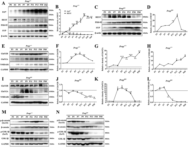
Fig. 1. Western blot (WB) analyses of PrPC (in wild-type (WT) only), REST, PSD-95, SYP, GluN2B, GluN2A, NMDAR1, total and phosphorylated β-catenin and GSK3β during WT Prnp (Prnp+/+) and PrP-null (Prnp0/0) mice hippocampal postnatal development.
a, e, m WB results in whole hippocampal lysates of WT mice (n = 6). c, i, n WB results in whole hippocampal lysates of Prnp0/0 mice (n = 6). b Quantitative analyses of (a). d Quantitative analyses of (f). f–h Quantitative analyses of (e). j-l Quantitative analyses of i. Immunoblot density in b and d normalized to β-actin. Immunoblot density in f–h and j–l normalized to GAPDH. All values were normalized (dashed lines) relative to corresponding data at P3 in each group. Data are presented as means ± SD (n = 6). *P < 0.05; **P < 0.01; ***P < 0.001 vs corresponding data at P3

