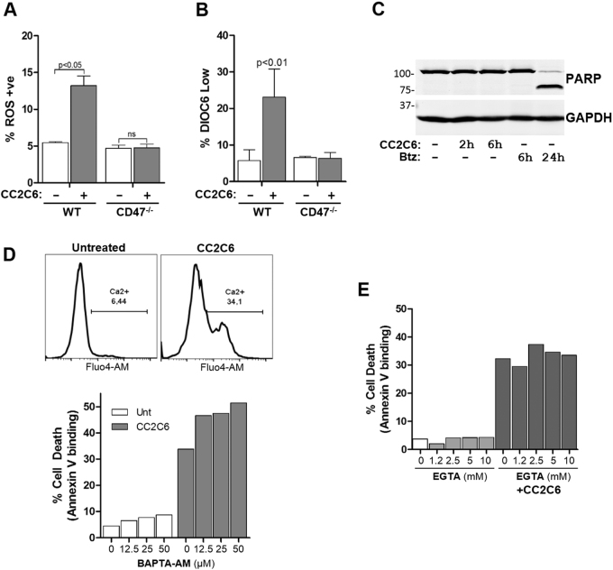
Fig. 4. Characterization of CC2C6-induced cell death.
a WT and CD47−/− Jurkat cells were treated with or without 125 ng/mL CC2C6 for 2 h and reactive oxygen species (ROS) generation assessed by flow cytometry using MitoSox labeling. b As in (a), loss of mitochondrial membrane potential (MMP) was assessed by flow cytometry using DiOC6 labeling. c Jurkat cells were untreated or treated with 125 ng/mL CC2C6 or bortezomib for the indicated times and cell lysates immunoblotted for PARP and GAPDH. d Top panel: Jurkat cells labelled with Fluo4-AM were treated with and without 125 ng/mL CC2C6 for 2 h and intracellular Ca2+ assessed by flow cytometry. Bottom panel: Jurkat cells were untreated or pretreated with BAPTA-AM at the indicated concentrations, followed by treatment with and without 125 ng/mL CC2C6 for 2 h. Cell death was assayed by flow cytometry using Annexin V binding. e As in (d), Jurkat cells were incubated with and without 125 ng/mL CC2C6 and EGTA at the indicated concentrations, and cell death assessed by flow cytometry. Error bars represent the standard deviation for n = 3 replicates. Representative of three independent experiments

