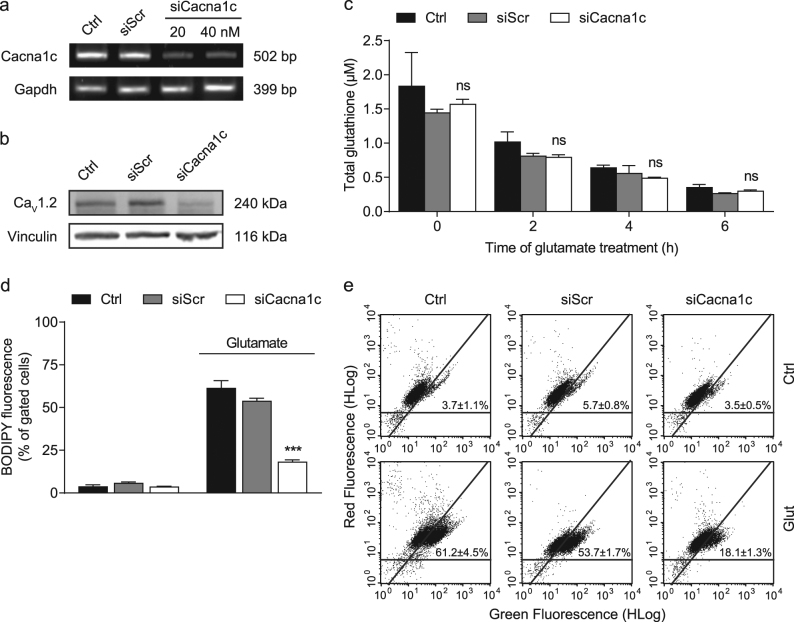
Fig. 1. siRNA-mediated knockdown of Cacna1c prevented lipid peroxidation, but not glutathione depletion following glutamate exposure.
a Cacna1c mRNA levels were analyzed 24 h after siRNA transfection with 20 and 40 nM. Gapdh served as internal control. b Protein samples were collected 48 h after transfection with 40 nM siRNA and the CaV1.2 expression levels were then identified by Western blot. Vinculin was used as loading control. c Total glutathione levels were calculated from three replicates per condition after 0, 2, 4, and 6 h of glutamate treatment (10 mM). Data are provided as mean + SD. d, e Lipid peroxidation in HT22 cells was determined using BODIPY staining after an 8-h incubation with 9 mM glutamate. The dot plots show representative replicates and the bar graph summarizes the associated experiment where three replicates per sample are shown as percentage of cells in the upper right quarter (mean + SD; 10,000 cells per replicate). Ctrl control, siScr scrambled siRNA, siCacna1c Cacna1c siRNA, Glut glutamate. ***p < 0.001; ns (not significant) compared to glutamate-treated ctrl (ANOVA, Scheffé’s-test)

