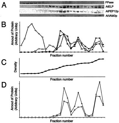
Figure 4.
AtVAM3p co-fractionates with PVC markers in Suc density gradients. A, Microsomal extracts of wild-type Arabidopsis plants were separated on Suc density gradients. Twenty-four fractions were taken, TCA precipitated, resuspended in SDS sample buffer, and equal volumes separated by SDS-PAGE. Strips of these blots were probed with antisera specific to: H+-pyrophosphatase (PPase), AtELP, C-terminal specific AtPEP12p, or AtVAM3p. B, These blots were digitized on a flat-bed scanner, and densitometry was performed with imaging software. Shown is a quantification of each fraction (relative to the total amount of each protein loaded onto the gradient) for H+-pyrophosphatase (PPase, ⋄), AtELP (□), AtPEP12p (▴), and AtVAM3p (●). C, The density profile of this gradient was determined by refractometry and is virtually linear. D, Microsomal extracts of plants either expressing T7-AtPEP12 (□) or T7-AtVAM3 (▴) were separated on Suc density gradients as described for A, and quantified as described in B. The density profile of each gradient was similar to that shown in C.

