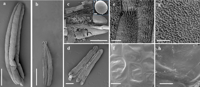
Fig. 2.
Appearance of the anther and pollen grain in the wild type (WT) and ms33-6029 mutant at stage 13 of anther development under scanning electron microscopy. a WT and b ms33-6029 anthers. c, c1, d, Pollen grains of WT (c, c1) and the absence of pollen grains in ms33-6029 (d). e, f The outermost surface of the epidermis of WT (e) and ms33-6029 (f) anthers. g, h The inner surface of the anther wall layers of WT (g) and ms33-6029 (h) anthers. Bars = 1 mm (a, b), 500 μm (c, d), 15 μm (c1), 10 μm (e, f), 5 μm (g, h)

