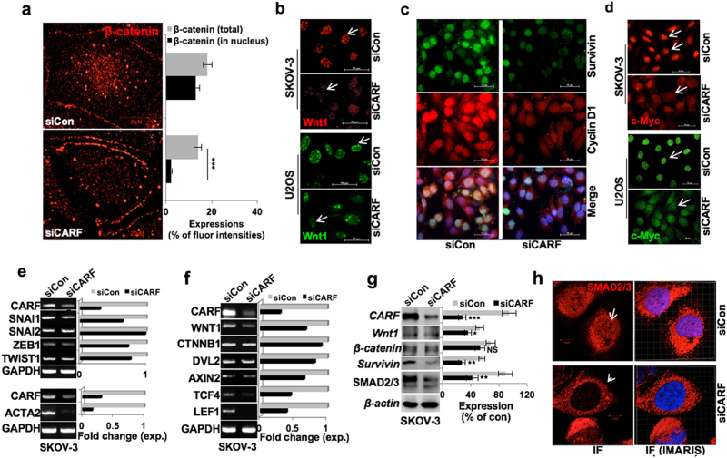
Fig. 6. CARF knockdown diminished EMT progression via abrogating β-catenin nuclear translocation.
a Immunostaining showing depletion of β-catenin expression in nucleus in CARF knockdown cells; quantitation of total and its nuclear level is shown at right. b Wnt1 immunostaining showing reduced nuclear foci in both, SKOV-3 and U2OS CARF-compromised cells. c Survivin and Cyclin D1 immunostaining showing their reduced levels in SKOV-3 CARF/siRNA cells. d c-Myc immunostaining showing a decrease in its expression in CARF knockdown both, SKOV-3 and U2OS cells. e Transcript levels of the SNAIL1, SNAIL2, ZEB1, TWIST1, and ACTA2 (αSMA), and f the β-catenin target genes; WNT1, CTNBB1, DVL2, AXIN2, TCF4, and LEF1 showed decrease in CARF knockdown cells; quantitation in fold change is shown on the right. g Immunoblots showing decrease in β-catenin and its effector proteins viz. Wnt1, survivin, and SMAD2/3 in SKOV-3 siCARF cells. h Immunostaining showing reduced SMAD2/3 nuclear localization in SKOV-3 siCARF cells; interactive IMARIS image with semi-transparent nuclear (blue) are shown on the right

