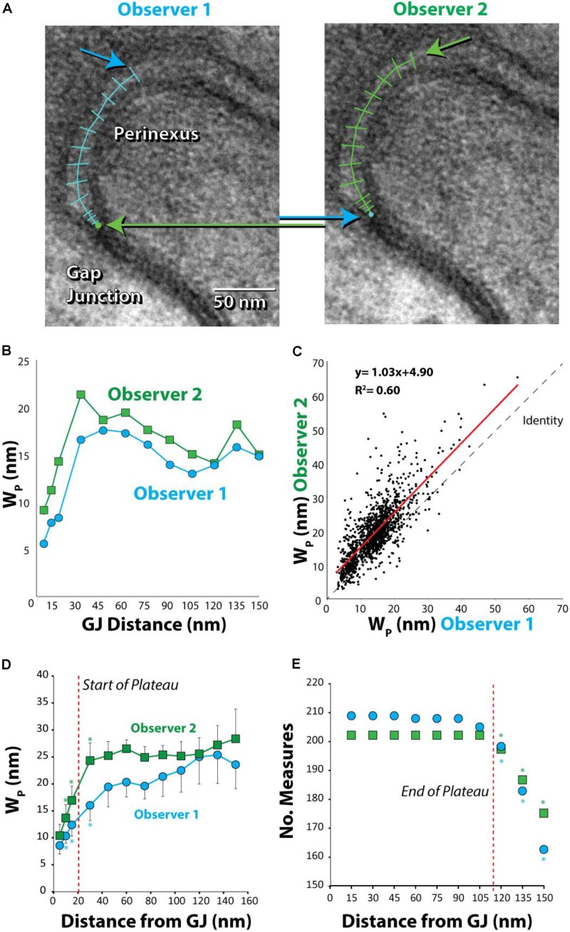
FIGURE 3.
Absolute, but not relative, perinexal widths differ between observers. (A) Representative TEM image from human left atrial appendage measured by two different observers. Arrows indicate different starting and ending points between observers. (B) Perinexal width (Wp) from (A) are different between observers 1 and 2. (C) Wp correlates between observers using data from the first 20 patients (149 images) but the y-intercept is different. (D) Wp changes from the gap junction edge up to 30 nm (∗p < 0.05). (E) Observers collect similar numbers of Wp measurements up to 105 nm from gap junction edge. (∗p < 0.05 relative to numbers at previous distance).

