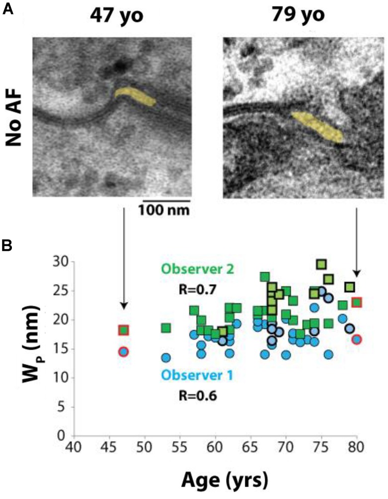
FIGURE 5.
Perinexal width correlates with age. (A) Representative TEM image from two patients without a history of atrial fibrillation, ages 47 and 79 years old (yo). (B) All blinded observers found a correlation by Spearman’s Rank correlation for all patient samples analyzed (Observer 1, R = 0.6. Observer 2, R = 0.7). Dark blue circles and dark green squares represent non-AF Wp values obtained by Observers 1 and 2, respectively, while bold-outlined light blue circles and bold-outlined light green squares represent AF Wp values obtained by Observers 1 and 2, respectively. Red circles and squares indicate samples represented by TEM images in (A).

