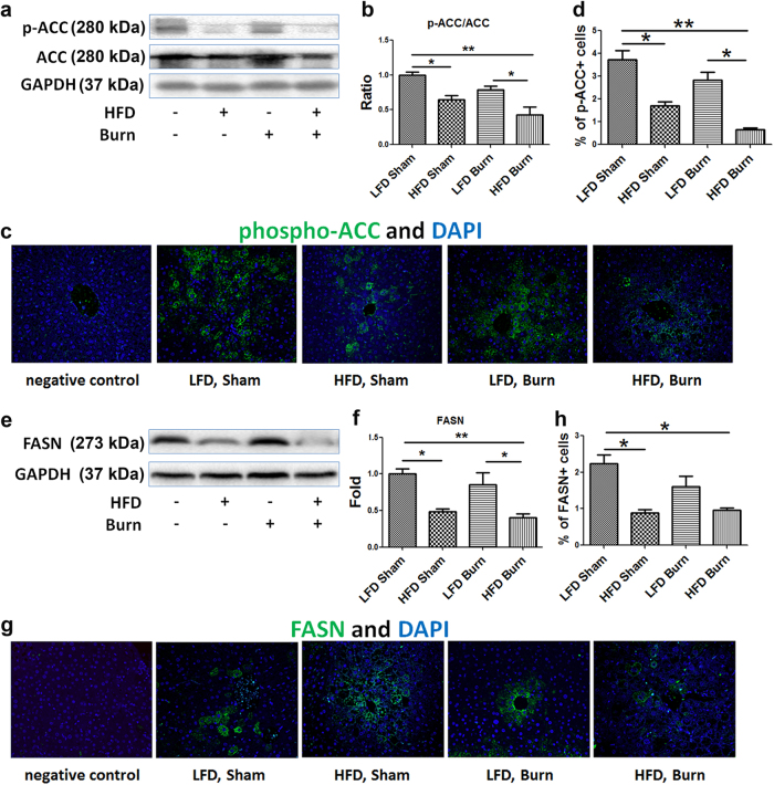
Fig. 2. Repression of de novo lipogenesis in HFD mice after thermal injury.
Representative images (a) and quantitative densitometric analyses (b) of the Western blot of phospho-ACC (Ser79) and ACC were presented alongside immunofluorescent staining of phospho-ACC (c, magnification ×200) and percentage of phospho-ACC positive cells (d) in liver tissue. Representative images (e) and quantitative densitometric analyses (f) of the western blot of FASN were presented alongside immunofluorescent staining of FASN (g, magnification ×200) and percentage of FASN positive cells (h) in liver tissue. Data are presented as means ± SEM. *P < 0.05 and **P < 0.01. N = 6 animals per group

