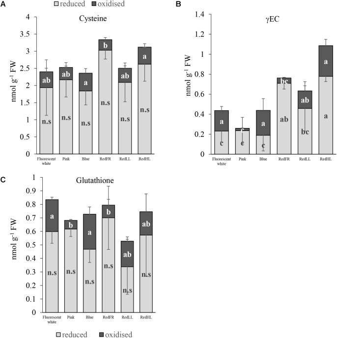
FIGURE 3.
Effects of light spectral distribution and light intensity on thiol levels and forms. (A) Cysteine. (B) γ-glutamylcysteine (γEC). (C) Glutathione. Values are the mean ± SD of 5 biological replicates per light treatment. The different letters indicate statistically significant differences at P < 0.05, using Tukey’s post hoc test.

