
FIGURE 5.
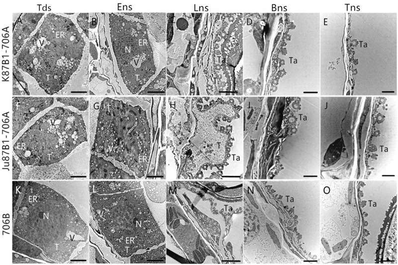
Transmission electron micrographs of the anthers in K87B1-706A (A–E), Ju87B1-706A (F–J), and 706B (K–O) during different developmental stages. Tds, tetrad stage (A,F,K); Ens, early uninucleate stage (B,G,L); Lns, late uninucleate stage (C,H,M); Bns, binucleate stage (D,I,N); and Tns, trinucleate stage (E,J,O). ER, endoplasmic reticulum; N, nucleus; T, tapetum; Ta, tapetosome; V, vacuole. Scale bars = 2 μm (A–O).