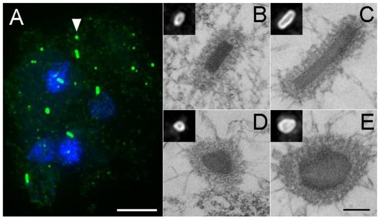
Figure 6.
Aberrant centrosome formation. Panel (A) shows a tetranucleated cell with supernumerary centrosomes (green, DdCP224 staining, DdKif9 null background). The arrowhead highlights the normal appearance for an interphase centrosome. Note here the number, distribution, and variability in centrosome sizes, as well as the lack of nuclear association. Scale bar = 5 μm. Panels (B–E) show thin section electron micrographs of selected centrosomes. Scale bar = 250 nm (B,D) present side and top views of wild type centrosomes, (C,E) show elongated versions. Insets show the same centrosomes by light microscopy. Panels (B,D,E) adapted with permission from Ref [29].

