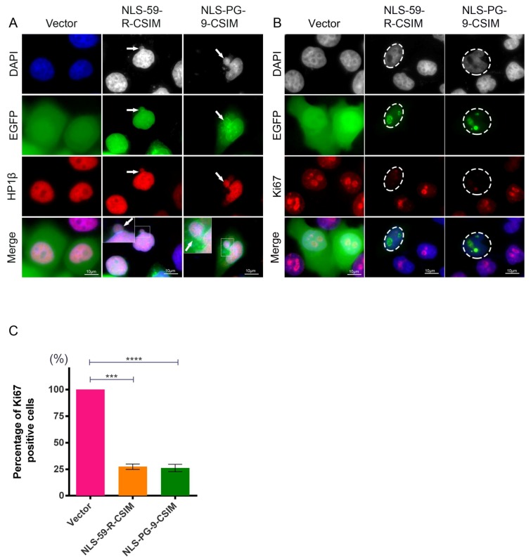
Figure 4.
Determination of the heterochromatin organization and cell proliferation activity. (A) Heterochromatin was stained with an anti-HP1β antibody. Representative images of HP1β staining in EGFP vector-, EGFP–NLS-59-R-CSIM-, and EGFP–NLS-PG-9-CSIM-transfected cells. Cytoplasmic HP1β staining shows the disorganized heterochromatin signals in EGFP–NLS-59-R-CSIM- and EGFP–NLS-PG-9-CSIM-transfected cells, as indicated by the arrowheads. Higher magnification images show the colocalization of DAPI and heterochromatin staining in the cytoplasm. Scale bar, 10 µm; (B) Ki67 was used to detect the actively replicating chromosomal DNA. Representative images of Ki67 staining and signals for EGFP fusion proteins are shown. Ki67 signals are barely detectable in EGFP–NLS-59-R-CSIM- and NLS-PG-9-CSIM-expressing cells, as indicated with circles; (C) Percentages of cells expressing both EGFP and Ki67. Nine hundred sixty cells expressing the EGFP vector, 919 cells expressing EGFP–NLS-59-R-CSIM, and 934 cells expressing EGFP–NLS-PG-9-CSIM were counted (n = 3). Scale bar, 10 µm.

