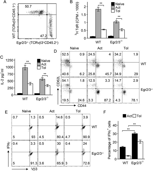
Figure 5.

Egr2/3 intrinsically control inflammatory activation of tolerant T cells in vivo. An equal number of bone marrow cells from wild‐type (WT) and CD2‐Egr2/3−/− (Egr2/3−/−) mice were adoptively transferred into irradiated wild‐type recipients. Six weeks after bone marrow reconstitution, wild‐type (CD45.1) and Egr2/3−/− (CD45.2) cells among gated CD4+TCRVβ3+ cells were quantified (A). Recipient mice were injected with SEA once to activate T cells (Act) or five times with 4 day intervals to induce tolerance (Tol). Twenty‐four hours after the last injection, CD4+TCRVβ3+CD45.1+ (WT) and CD4+TCRVβ3+CD45.2+ (Egr2/3−/−) cells were isolated and re‐stimulated with SEA loaded dendritic cells in vitro. (B) Proliferation was measured 3 days after re‐stimulation. (C) IL2 in supernatants was measured by ELISA 24 h after re‐stimulation. (D) CD62L and CD44 expression on gated CD4+TCRVβ3+CD45.1+ (WT) and CD4+TCRVβ3+CD45.2+ (Egr2/3−/−) cells from recipient mice was analyzed 24 h after the last SEA injection. (E and F) IFNγ producing cells among gated CD4+TCRVβ3+ cells were analyzed after re‐stimulation for 24 h. Data in A, D, E are from five mice in each group and are representative of two experiments. Data in B, C and F are from five mice for each group and are representative of two experiments. Data in B, C, and F are the mean ± s.e.m. and were analyzed with Kruskal–Wallis tests followed by Conover tests with Benjamini–Hochberg correction. N.S. not significant, *p < 0.05, **p < 0.01.
