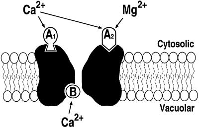
Figure 8.
Simplified model for the regulation of SV channels by cytosolic and luminal Ca2+ and Mg2+ in fava bean guard cells. A1, High-affinity Ca2+-binding site on the cytosolic side, which is not activated by Mg2+. A2, Low-affinity binding site on the cytosolic side, which can be occupied by either Mg2+ or Ca2+. B, Vacuolar Ca2+-binding site, which is not affected by vacuolar Mg2+. For the activation of SV channels, both activation sites A1 and A2 need to be occupied (see “Discussion”). The cytosolic and vacuolar membrane sides are labeled.

