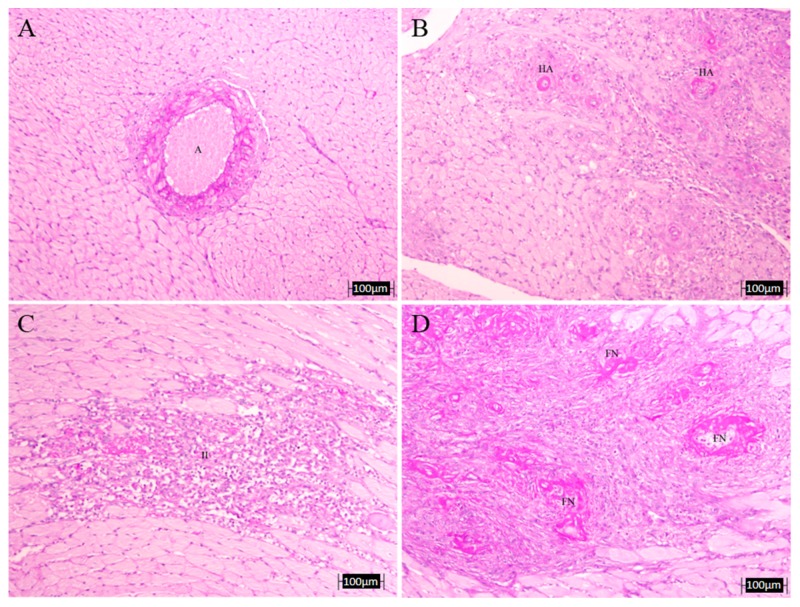
Figure 6.
Representative microphotographs of heart lesions. Upper left: intramyocardial artery (A) of a control non-treated rat (no morphological alterations, PAS, 10×); (B) hyaline arteriopathy (HA) in a coronary artery of L-NAME-treated rat (PAS, 10×); (C) intense vascular damage with inflammatory infiltrate (II) and myocardiocytes lesions in a L-NAME-treated rat heart (PAS, 10×); (D) fibrinoid necrosis (FN) in a coronary artery of L-NAME-treated rat (PAS, 10×).

