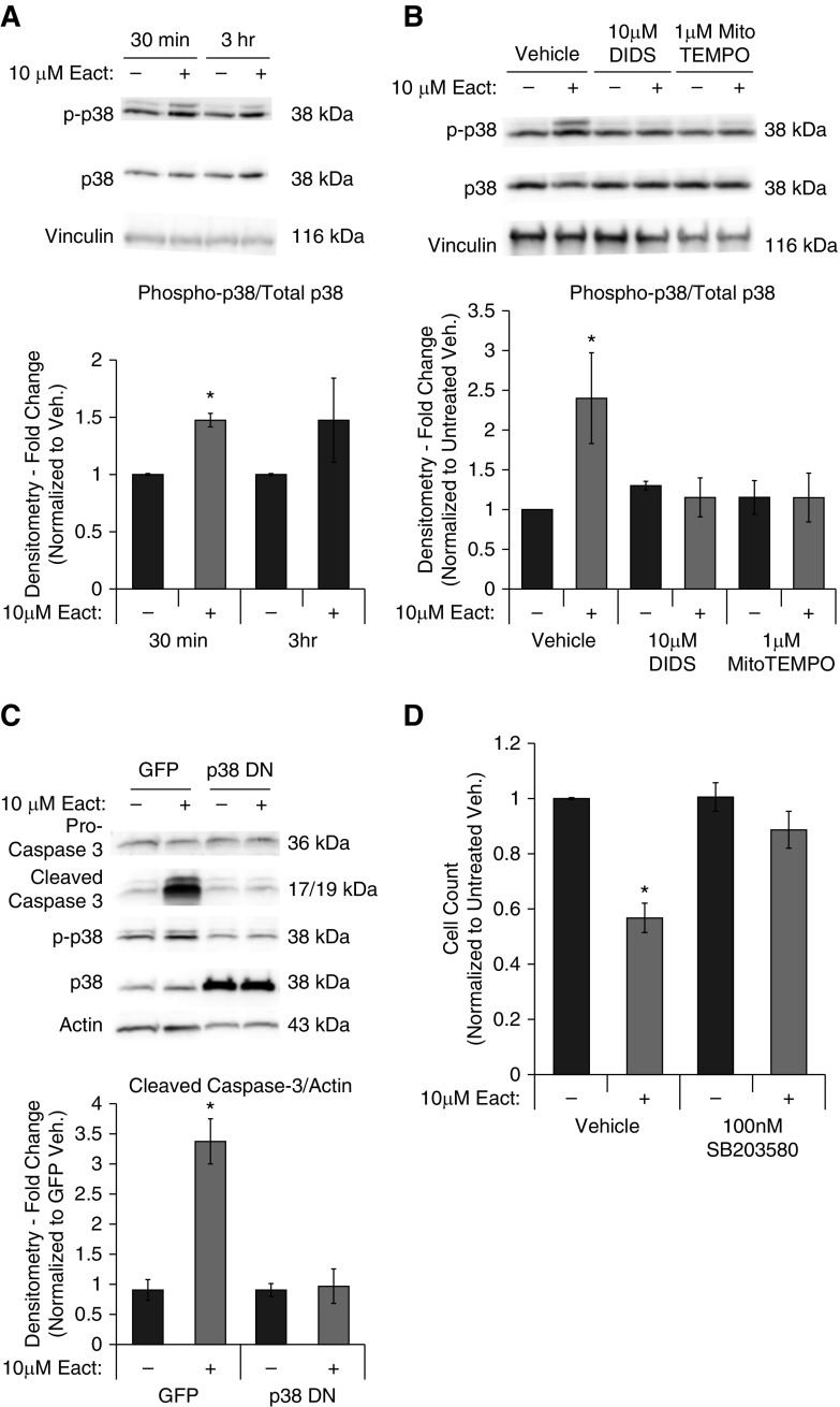
Figure 4.
Ano1 activation is p38–mitogen-activated protein kinase (MAPK) dependent. RLMVECs were seeded subconfluently and allowed to adhere and infected with virus, as indicated. Cells were then quiesced for 24 hours followed by drug treatments in complete media. (A) p38 phosphorylation in response to Eact treatment was assessed by immunoblot at both 30-minute and 3-hour time points (n = 5, ANOVA with Tukey multiple comparison, *P < 0.05). (B) RLMVECs were pretreated with either 10 μM DIDS, 1 μM Mito-TEMPO, or vehicle for 1 hour and then treated with vehicle or 10 μM Eact for 30 minutes. p38 phosphorylation was then evaluated by immunoblot (n = 5, ANOVA with Tukey multiple comparison, *P < 0.05). (C) Cells were infected with adenoviruses containing GFP or dominant-negative p38 constructs and pro-caspase 3, cleaved caspase-3, phospho-p38, total p38, and actin expression was assessed using immunoblot (n = 4, ANOVA with Tukey multiple comparison, *P < 0.05). (D) Cells were treated with p38-MAPK inhibition with vehicle or 10 μM Eact in the presence/absence of SB203580 for 24 hours and cell counts were performed (n = 4, ANOVA with Tukey multiple comparison, *P < 0.05). Data are presented as mean (±SEM). Readers may view the uncut gel for Figure 4C in the data supplement. DN = dominant negative.

