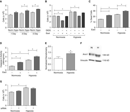
Figure 7.
Hypoxia effects on RLMVECs. Cells were seeded subconfluently and then quiesced with reduced serum MCDB-131 media for 24 hours. At the start of the experiments, media were switched over to complete MCDB and cells were either exposed to normoxia (Norm) or hypoxia (Hypo) for the indicated time. (A) RLMVECs exposed to Hypo (1% FiO2) had higher cell counts than their normoxic counterparts (n = 4–6, *P < 0.05 versus Norm of time point). (B) RLMVECs were exposed to either normoxia or hypoxia for 3 days and pretreated with vehicle/DIDS before treatment with vehicle/10 μM Eact for an additional 24 hours, followed by cell counts (n = 4, *P < 0.05 using three-way ANOVA followed by Tukey post hoc pairwise comparison). Apoptosis in response to hypoxia and/or Eact was assessed by annexin-V staining (C) and caspase-3 activity (D) (n = 5, *P < 0.05, two-way ANOVA). Ano1 expression was upregulated in response to exposure to 3 days of hypoxia (E) (n = 15, *P < 0.05). (F) A representative blot is shown. (G) In another series of experiments, cells were transfected with either 100 nM scrambled or Ano1 siRNA in serum reduced media overnight. Media were then switched to complete MCDB media, and cells were placed into either normoxia or hypoxia for 24 hours and assessed by cell counts (n = 6, *P < 0.05, two-way ANOVA). All data are presented as mean (±SEM). Readers may view the uncut gel for Figure 7F in the data supplement.

