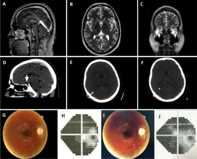
Figure 1.

Imaging, fundoscopic pictures and visual field of the patient A) Sagittal T2-weighted MRI. B) Unremarkable axial T2-weighted MRI. C) Coronal T2-weighted MRI image demonstrates bilateral widening of the optic nerve sheath. D) CT venogram of the patient showing a partially empty sella. E, F) CT of the brain, 1 day postoperatively, demonstrating VP shunt insertion. G) Fundoscopic picture showing improved optic disc edema in the patient’s right eye (3 months postoperatively). H) Humphrey visual field test showing residual constriction of the patient’s peripheral vision in the right eye (3 months postoperatively). I) Fundoscopic picture of the patient’s right eye (7 months postoperatively). J) Humphrey visual field test showing some improvement of the patient’s peripheral vision in the right eye (7 months postoperatively).
