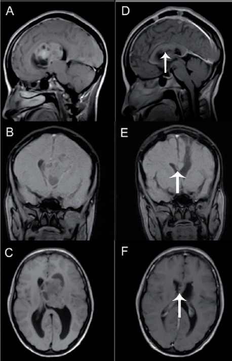
Figure 1.

A 30-year-old woman presenting with a headache, blurry vision, and attacks of generalized body numbness for 2 months. A-C) Sagittal, coronal, and axial T1 Magnetic Resonance Imaging (MRI) with and without contrast enhancement showing third ventricular tumor with extension to the left lateral ventricle. She underwent left frontal craniotomy, frontal transcortical approach with subtotal excision of the tumor. The pathology revealed central neurocytoma. D-F) Postoperative with and without contrast enhancement T1 sagittal, coronal, and axial scans showing small residual tumor (arrow) under the corpus callosum. She postoperatively developed right side hemiparesis. In her follow-up, 6 and 9 months following surgery, her weakness improved significantly. We think the reason for her weakness is due to the retraction on the motor control areas.
